
Figure 7.
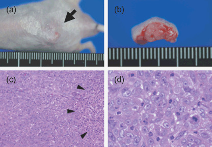
Heterotransplantation of ThyL‐6 cells into a nude mouse. (a) A representative view of tumor (arrow) in the back of a nude mouse. (b) Cross‐section of subcutaneous tumor mass lesion in a nude mouse. (c) A microscopic examination of the tumor produced in a nude mouse (low power magnification view) with hematoxylin and eosin staining. Tumor cells had proliferated with small amount of interstitial fibrous connective tissue. Liquefactive necrotic tissue was seen in the tumor mass (arrow head). (d) High power magnification view of the established tumor. Polymorphic cells with round or oval nuclei and a large round nucleolus had proliferated by mitosis, and neutrophils had infiltrated into the tumor nests.