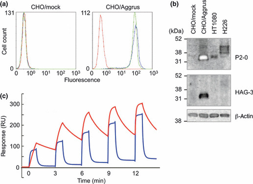
Figure 1.

Characterization of two novel anti‐human Aggrus mAb, P2‐0 and HAG‐3. (a) Stably‐transfected CHO/mock and CHO/Aggrus were treated with control mouse IgG1 (control IgG) ( ), P2‐0 (
 ), and HAG‐3 (
 ). After incubation with the second antibody, Aggrus expression was analyzed by flow cytometry. (b) Cells were lysed and immunoblotted with the indicated antibodies. (c) Interaction between the human Aggrus protein and anti‐human Aggrus mAb was estimated by surface plasmon resonance analysis. Five different concentrations of antibodies (P2‐0, 6.25–100 nM; HAG‐3, 62.5–1000 nM) were passed over a sensor chip with the immobilized Aggrus protein for 1 min before the flow was switched to the buffer alone for another 2 min in a single cycle. Equilibrium dissociation constants (K
D) are shown. P2‐0 (K
D = 9.30 x 10⁻⁹ M) (
 ) and HAG‐3 (K
D = 4.30 x 10⁻⁷ M) (
 ).
).
