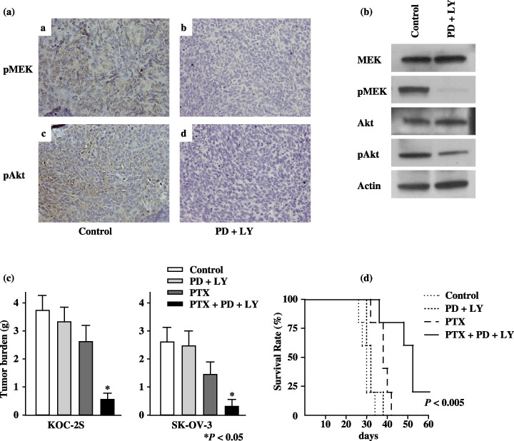
Figure 5.

Treatment with paclitaxel (PTX) combined with both mitogen‐activated protein kinase kinase (MEK) and phosphatidylinositol 3′‐kinase (PI3K) inhibitors prolongs survival in mice with implanted KOC‐2S cells. (a) Immunohistochemical stains of representative tumor tissue samples from mice implanted with KOC‐2S cells and treated with 7 mg/kg PD98059 (PD) and 25 mg/kg LY294002 (LY). The brown staining at the upper and lower left indicates the phosphorylated (p) MEK and pAkt proteins, respectively. The results shown represent duplicate experiments. (b) The levels of pMEK and pAkt protein were determined 24 h after intravenous treatment with phosphate‐buffered saline (PBS), or PD and LY by western blotting. Those proteins were effectively downregulated only in tumors from mice treated with PD and LY. The results shown represent duplicate experiments. (c) Female nude mice (five per group) were given an intraperitoneal (i.p.) injection of 2 × 10⁶ KOC‐2S cells or SK‐OV‐3 cells followed by weekly i.p. injections of 25 mg/kg PTX, and/or 7 mg/kg PD and 25 mg/kg LY for 4 weeks. Tumors were collected and weighed on day 28. In KOC‐2S cell‐ and SK‐OV‐3 cell‐inoculated mice, the weight of the peritoneally disseminated tumors was significantly lower in the mice treated with PTX combined with PD and LY than with the other treatments (P < 0.05). (d) Female nude mice (5 per group) were given an intraperitoneal (i.p.) injection of 2 × 10⁶ KOC‐2S cells followed by weekly i.p. injections of 25 mg/kg PTX, and/or 7 mg/kg PD and 25 mg/kg LY for 4 weeks. Treatment with PTX, PD, and LY prolonged survival relative to treatment with PBS, PD and LY, or PTX (P < 0.005).
