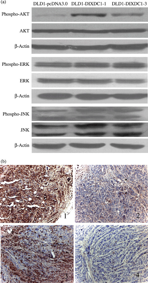
Figure 4.

Overexpression of DIXDC1 activated the PI3K/Akt pathway in DLD1 cells. (a) Cell lysates were subjected to immunoblotting using antibodies against Akt, p‐Akt, ERK, p‐ERK, JNK, and p‐JNK. (b) Immunohistochemical staining on DLD1 xenograph tumors. Tumors from DLD1‐DIXDC1‐1‐injected mice showed strong cytoplasmic DIXDC1 (Fig. 4b‐1) and (p)‐Akt staining (Fig. 4b‐2), compared with tumors from DLD1‐pcDNA3.0‐injected mice (Figs 4b‐3 and 4b‐4).
