
Figure 3.
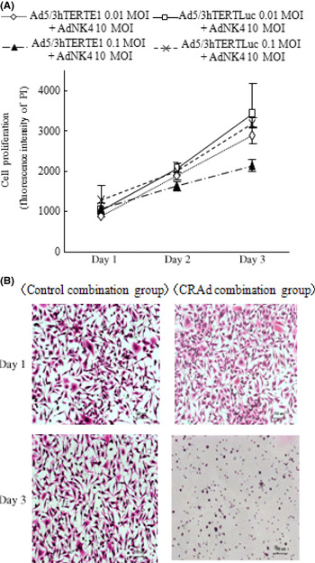
Ad5/3hTERTE1 enhanced the inhibitory effects of Ad‐NK4 on cancer cell invasion. (A) SUIT‐2 cells were infected with Ad‐NK4 (10 MOI) and either Ad5/3hTERTE1 or Ad5/3hTERTLuc (0.01 MOI or 0.1 MOI). Cell proliferation was measured by the fluorescence intensity of propidium iodide (PI), which correlates with the cell number, on the indicated days. All experiments were performed in triplicate wells and repeated at least three times, and each value represents the mean ± SD of three independent samples. (B) Control‐combination group SUIT‐2 cells were treated with Ad‐NK4 (10 MOI) and Ad5/3hTERTLuc (0.01 MOI), and the culture media were collected. Fresh untreated SUIT‐2 cells were seeded in DMEM mixed with conditioned culture media in the inner chamber and cultured with conditioned culture media in the outer chamber. After 48 h of culture in the presence of 3 ng/mL hepatocyte growth factor (HGF), cells that had invaded the lower surface of the Matrigel‐coated membrane were fixed with 70% ethanol, and stained with H&E. Photomicrographs of SUIT‐2 cells that invaded to the lower surface of the Matrigel‐coated membrane are shown.