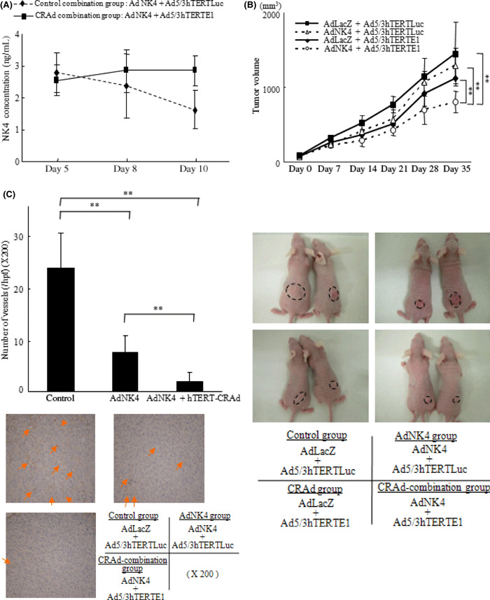
Figure 4.

Combination with Ad5/3hTERTE1 sustained the expression levels of NK4 delivered by Ad‐NK4 in tumors and enhanced the therapeutic efficacy in vivo. Six‐week‐old female nude mice were used in the in vivo study. (A) Mice were injected subcutaneously with SUIT‐2 cells. The control‐combination group mice were administered Ad‐NK4 and Ad5/3hTERTLuc peritumorally, and the CRAd‐combination group mice were administered Ad‐NK4 and Ad5/3hTERTE1 on day 1. Five mice in each group were sacrificed and their tumors were excised for protein extraction on days 5, 8, and 10. NK4 was measured by ELISA. Each value represents the mean ± SD of three independent samples. **P < 0.01. (B) Seven mice in each group were subcutaneously implanted with SUIT‐2 cells in the back. Each group was administered weekly peritumoral injections on days 1, 8, and 15 as follows: control group, Ad‐LacZ+Ad5/3hTERTLuc; Ad‐NK4 group, AdAd‐NK4 + Ad5/3hTERTLuc; CRAd group, Ad‐LacZ + Ad5/3hTERTEl; and CRAd‐combination group, Ad‐NK4 + Ad5/3hTERTEl. The size of the tumors was measured weekly, and the volume of the tumors was calculated. (C) Five mice in each group (control group, Ad‐LacZ + Ad5/3hTERTLuc; Ad‐NK4 group, AdAd‐NK4 + Ad5/3hTERTLuc; and CRAd‐combination group, Ad‐NK4 + Ad5/3hTERTEl) were treated the same way, and on day 21, tumors were excised for immunohistochemical staining. Microvessels were determined by anti‐CD31 antibody and the microvessel density (MVD) was counted. Arrows indicate microvessels. Data are expressed as mean ± SD. **P < 0.01.
