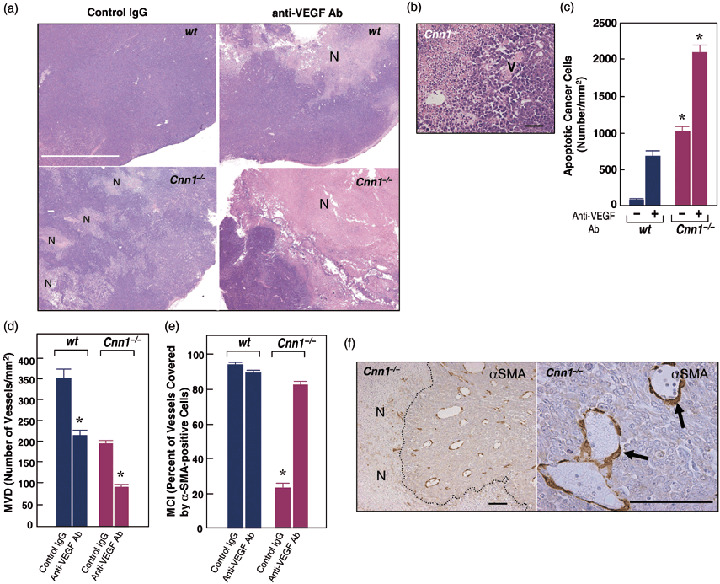
Figure 4.

Deletion of smooth muscle calponin (Cnn1) –/– leads to sensitization of cancer cells to anti‐vascular endothelial growth factor (VEGF) antibody treatment. (a) Macroscopic view of hematoxylin–eosin (H&E)‐stained sections of Lewis lung carcinoma (LLC) tumors 18 days after initial anti‐angiogenesis therapy showed the most extensive necrosis of cancer cells (N) in anti‐VEGF antibody‐treated Cnn1 –/– mice. Note that in Cnn1 –/– mice, even non‐treated tumor showed focal necrosis of cancer cells (N). White bar = 5 mm. (b) Higher magnification pictures show prominent destruction of tumor vasculature (V) in anti‐VEGF antibody‐treated LLC tumors in Cnn1 –/– mice. Black bar = 50 µm. (c) Quantitation of TUNEL assay of tissue sections from the tumors in part (a), showing a striking increase in the number of apoptotic cancer cells in anti‐VEGF antibody‐treated LLC tumors in Cnn1 –/– mice. Mean ± SE (30 high‐power field analyses from four mice). *P < 0.05. (d–f) Treatment with anti‐VEGF antibody significantly reduced the microvessel densities (MVD) in both wild‐type and Cnn1 –/– mice (d) and normalized the pericyte coverage index (PCI) in Cnn1 –/– mice (e). Blood vessels covered by smooth muscle α‐actin (α‐SMA)‐positive mural cells in Cnn1 –/– mice were refractory to the anti‐VEGF antibody therapy (f). Scale bar = 100 µm.
