
Figure 2.
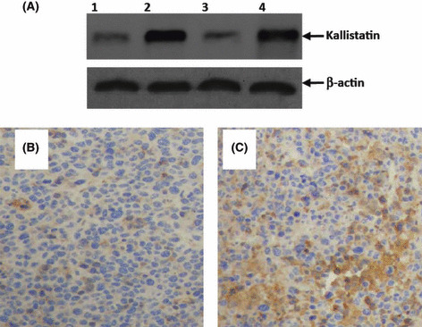
Intense expression of kallistatin in situ after intratumoral gene transfer. (A) Homogenates of tumors from mice treated with pcDNA3.1 (lane 1), Kalli‐pcDNA3.1 (lane 2), meloxicam (lane 3), or Kalli‐pcDNA3.1 + meloxicam (lane 4) one week earlier underwent Western blot analysis with either anti‐kallistatin or β‐actin Abs. Illustrated are representative tumor sections prepared one week following intratumoral gene transfer of pcDNA3.1 (B) and Kalli‐pcDNA3.1 (C) plasmids. The sections were stained with an anti‐kallistatin Ab.