Figure 4.

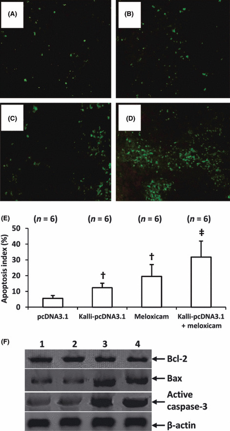
Combining kallistatin gene and meloxicam induces more apoptotic cells. Representative tumor sections prepared from mice treated with pcDNA3.1 (control) (A), Kalli‐pcDNA3.1 (B), meloxicam (C), or Kalli‐pcDNA3.1 + meloxicam (D) one week earlier. Tumor sections were stained with the TUNEL agent to view apoptotic cells. (E) TUNEL‐positive cells were counted to calculate the apoptosis index. †Significant difference in the apoptosis index from control. ‡Highly significant difference in the apoptosis index from control. n, number of tumors assessed. (F) Tumors from mice treated with pcDNA3.1 (lane 1), Kalli‐pcDNA3.1 (lane 2), meloxicam (lane 3), or Kalli‐pcDNA3.1 + meloxicam (lane 4), were homogenized and subjected to Western blot analysis to detect expression of Bcl‐2, Bax, and active caspase‐3. β‐actin served as an internal control.
