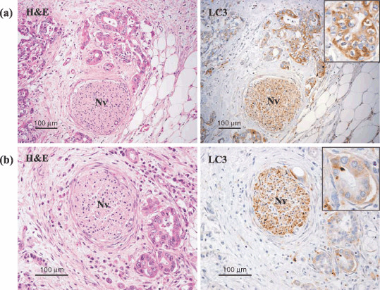
Figure 2.

LC3 protein was detected in pancreatic cancer tissue by immunohistochemical staining with LC3 antibody. Cancer tissue stained immunohistochemically with LC3 antibody and corresponding hematoxylin–eosin (HE)‐stained sections are shown. A nerve cell (Nv) was used as an internal positive control to validate immunohistocheimical staining and evaluate the level of intensity of LC3 expression. The inserts are the photographs at higher magnification. (a) The cancer cells that stained as or more intensely for LC3 than the nerve cells were recorded as strongly positive. (b) The cancer cells that stained less intensely for LC3 than the nerve cells were recorded as weakly positive.
