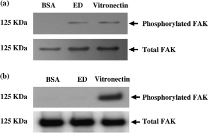
Figure 6.

Tyrosine phosphorylation of focal adhesion kinase (FAK). Lewis lung cancer (LLC) and KLN205 cells were placed for 45 min at 37°C on dishes that had been coated with endostatin (ED; 50 µg/mL), vitronectin (20 µg/mL) or bovine serum albumin (BSA; 10 mg/mL). Cell lysates containing equal amounts of protein were immunoprecipitated with anti‐FAK antibody, and one‐half of the precipitates was analyzed by immunoblotting with antiphosphotyrosine antibodies (top panel). The other half was probed with anti‐FAK antibody to confirm loading (bottom panel). Note that high levels of phosphorylated FAK in LLC cells plated on ED and vitronectin were observed (a). In contrast, phosphorylation of FAK was not observed in KLN205 cells cultured on ED (b).
