
Figure 3.
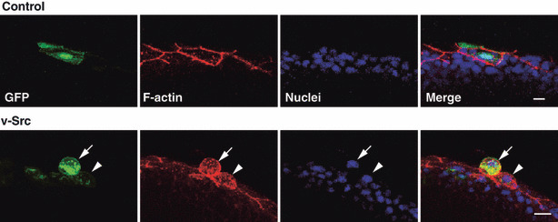
Apical extrusion of a v‐Src‐expressing cell from a monolayer of the enveloping layer in a zebrafish embryo. Immunofluorescence images (semilateral view) of zebrafish embryos (at 8–9 h postfertilization), injected with the GFP‐ (control) or v‐Src‐expressing vector at the 1‐ to 2‐cell stage. Embryos are stained with phalloidin (red) and Hoechst (blue). Arrowhead and arrow indicate the v‐Src‐expressing cell with increased cell height and extruded v‐Src‐expressing cell, respectively. Scale bar = 10 μm. (Adapted from Kajita et al., 5 with permission, fig. 3.)