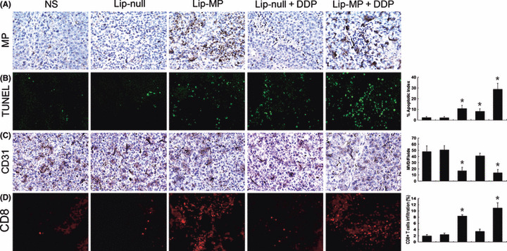
Figure 4.

Histochemical analysis of tumors. (A) Immuohistochemical analysis of vesicular stomatitis virus matrix protein (VSV‐MP) expression in tumor tissue. VSV‐MP‐expressing cells could be detected in both Lip‐MP and Lip‐MP + CDDP groups after intravenous administration of pVAX‐MP:lipo complexes versus the other groups. (B) Apoptotic tumor cells within tumor tissues were evaluated by TUNEL assays. An apparent increase in the number of apoptotic cells and apoptotic index was observed within the tumor tissues treated with Lip‐MP + CDDP compared with the control groups (P < 0.05). Data represent the mean apoptotic index ± SDs of cancer cells. (C) Inhibition of angiogenesis assayed by immunohistochemistry with CD31. The number of vessels per ×200 field was counted as described in the Materials and Methods. The Lip‐MP + CDDP‐ and Lip‐MP‐treated groups displayed decreased microvessel density as compared with the control groups (P < 0.05). (D) Fluorescence staining of infiltrated lymphocytes. The frozen sections were stained with anti‐CD8+ monoclonal antibodies as described in the Materials and Methods. CD8+ cytotoxic lymphocyte infiltration was significantly enhanced in the tumor tissues of Lip‐MP + CDDP‐ or Lip‐MP‐treated groups. The number of infiltrating CD8+ cytotoxic lymphocyte was further quantified by flow cytometry. Original magnification, ×200. The columns in all graphs correspond to the labeled columns in the picture. * P < 0.05, significantly different from the normal saline (NS) or Lip‐null groups.
