
Figure 6.
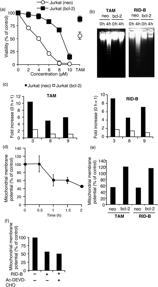
Mitochondria are involved in ridaifen (RID)‐B‐induced apoptosis. (a) Jurkat (neo) and Jurkat (bcl‐2) cells were incubated with the indicated doses of RID‐B or IC50 value (30 µM) of tamoxifen (TAM) (control) for 4 h. Cell viability was observed with a WST‐8 assay. The results are presented as a comparison with the non‐additive control. Each bar denotes the standard deviation (n = 3). (b) Jurkat (neo) and Jurkat (bcl‐2) cells were incubated with or without 4 µM RID‐B or IC50 value of TAM (for control) for 4 h. DNA fragmentation was carried out by agarose gel electrophoresis. (c) Jurkat (neo) and Jurkat (bcl‐2) cells were incubated with 4 µM RID‐B or IC50 value of TAM (for control) for 4 h. Caspase‐3, ‐8, and ‐9 assays were carried out. The results are presented as a comparison with the non‐additive control for each of the cells and assayed caspase numbers only are indicated in the graph. Data are representative of three independent experiments. (d) The mitochondrial membrane potential was observed by 5,5′,6,6′‐tetrachloro‐1,1′,3,3′‐tetraethylbenzimidazolylcarbocyanine iodide (JC‐1). Jurkat (neo) cells were incubated with 4 µM RID‐B for the indicated times and stained with JC‐1. The results are presented as a comparison with the non‐additive control. Each bar denotes the standard deviation (n = 3). (e) Jurkat (neo) and Jurkat (bcl‐2) cells were incubated with 4 µM RID‐B or IC50 value of TAM (for control) for 2.5 h and stained with JC‐1. The results are presented as a comparison with the non‐additive control. Data are representative of three independent experiments. (f) Jurkat cells were incubated with or without 30 µM acteyl (Ac)‐DEVD‐CHO for 1 h, and thereafter incubated with or without 4 µM RID‐B for 4 h. Mitochondrial membrane potential was observed by JC‐1. The results are presented as a comparison with the non‐additive control. Data are representative of three independent experiments.