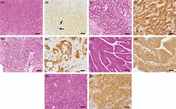
Figure 6.

Histopathology (A–E, using H&E stain) and immunohistochemistry for thioredoxin (Trx) (a–e, using the avidin–biotin complex method) in tissue‐arrayed normal and cholangiocarcinoma (ChC) human liver tissues. Trx were reliably overexpressed in human ChCs, irrespective of tumor type or neoplastic cell differentiation grade. Compare the weak expression of Trx in normal bile ducts (arrows). A and a, normal liver; B and b, well‐differentiated ChC; C and c, moderately differentiated ChC; D and d, papillary type ChC; E and e, poorly differentiated ChC. Bars = 25 μm.
