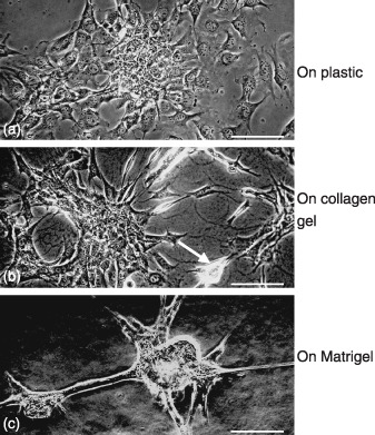
Figure 2.

Phase contast micrographs of GIF‐11 cells cultured on (a) plastic, (b) collagen gel, and (c) Matrigel for 1 day. (b) Some cells on the collagen gel attach closely to each other to form small cell masses (arrow), (c) whereas on Matrigel, much greater cell masses are formed. Scale bars = 100 µm.
