Figure 5.

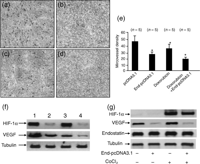
Endostatin gene transfer synergizes with doxorubicin to inhibit tumor angiogenesis. Illustrated are representative tumor sections prepared 2 weeks after treatment from mice receiving (a) pcDNA3.1 (control), (b) End‐pcDNA3.1, (c) doxorubicin, or (d) doxorubicin + End‐pcDNA3.1 treatment. (e) Tumor microvessels in sections were stained with the anti‐CD31 antibody and counted in blindly chosen random fields to record microvessel density. A significant difference in microvessel density between tumors treated with doxorubicin, or End‐pcDNA3.1 versus control is denoted by ‘*’, and a highly significant difference between tumors treated with combinational therapy with doxorubicin + End‐pcDNA3.1 and control by ‘**’. (f) Homogenates of tumors from mice treated with pcDNA3.1 (lane 1), End‐pcDNA3.1 (lane 2), doxorubicin (lane 3), or doxorubicin + End‐pcDNA3.1 (lane 4) were western blotted with anti‐ hypoxia‐inducible factor (HIF)‐1α (upper panel), ‐vascular endothelial growth factor (VEGF; middle panel) and ‐tubulin (lower panel) antibodies. (g) Western blot analysis of HIF‐1α, VEGF and endostatin expression in HepG2 cells in vitro. HepG2 cells were transfected with End‐pcDNA3.1, followed by CoCl2 treatment. Cell lysates were western blotted with anti‐HIF‐1α, VEGF, endostatin and tubulin antibodies.
