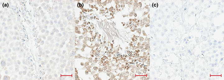
Figure 5.

Immunohistological analysis of seminiferous tubules sections: immunoperoxidase staining of p53. (a) p53RE‐Luc mouse before doxorubicin treatment; (b) p53RE‐Luc mouse after treatment and (c) control section with no primary antibody (magnification ×40). Bar: 100 micrometers.
