
Figure 1.
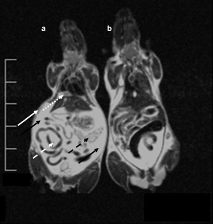
Magnetic resonance imaging (MRI) of disseminated tumor growth (ventro‐dorsal view). MRI carried out with a 1.5 Tesla MRT (Philips, the Netherlands) using a wrist coil. For the scan the 2D Turbospin Echo T2 program was chosen with a scan time of 11 min and a repetition time TR 2500 ms. (a) Mouse 3 weeks after tumor cell inoculation: the diaphragm is covered with a thick layer of tumor cells (dotted white arrow). Knotty metastases are visible at the caudal mesentery (dashed white arrow) and the peritoneal serous membrane (white arrow). The pancreas is heavily infiltrated with tumor cells (dashed black arrow). The peritoneal cavity is filled with ascites (black arrows). (b) Control mouse without tumor cells.