
Figure 4.
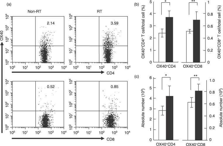
Irradiation augments OX40+ T cells in draining lymph nodes (DLN). Lewis lung carcinoma‐ovalbumin cells (1 × 10⁶) were inoculated intradermally into C57BL/6 mice, and irradiated or non‐irradiated on day 4. DLN obtained from both mice on day 8 were labeled with phycoerythrin‐conjugated anti‐OX40 mAb, followed by staining with fluorescein isothiocyanate‐conjugated anti‐CD4 or CD8 mAb. (a) Representative data from flow cytometric analysis. The numbers in each graph indicate the percentage of OX40+ cells in total cells from DLN. (b,c) Flow cytometric analysis pooled from five separate analyses. Data are presented as the mean ± SE. White and black bars indicate non‐irradiated mice (non‐radiotherapy (RT)) and irradiated mice (RT), respectively. (b) Proportion of OX40+ cells in DLN from the non‐RT and RT groups. *P = 0.002; **P = 0.02. (c) Absolute number of OX40+ cells in DLN from the non‐RT and RT groups. *P = 0.02; **P = 0.02.