
Figure 3.
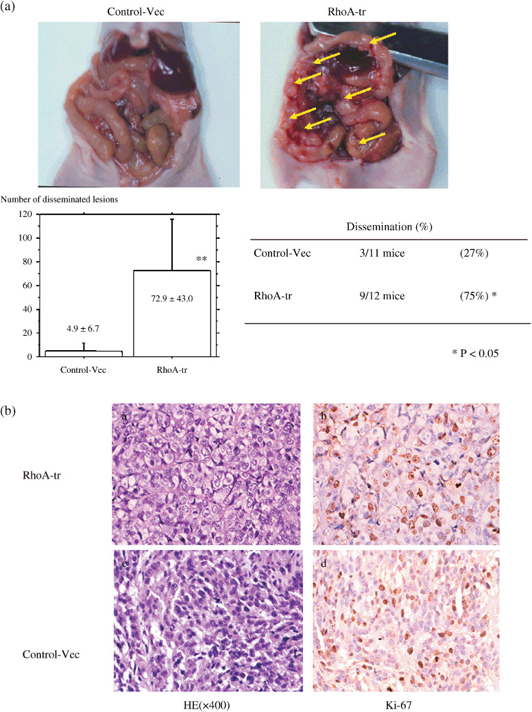
(a) Nude mouse tumorigenicity assay. RhoA‐transfected cells induced many small tumors 2 months after injection, whereas a small number of tumors developed in mice injected with vector‐transfected cells. The frequency of nude mouse dissemination of RhoA‐transfected SKOV3 cells was significantly elevated compared with vector cells after 2 months (P = 0.014). (b) Pathological analysis of tumors (Fig. 3a). Pathologically, tumors induced by RhoA‐transfected cells showed prominent nucleoli; however, nucleoli did not show a difference in Ki‐67 positivety in a histology examination of the sowing focus compared with the control, as for RhoA‐transfected cells (×400).