
Figure 8.
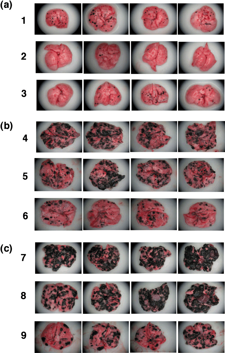
Macroscopic visualization of lung metastases. Metastatic spots on lung surfaces were observed with zoom stereo microscope at ×10 magnification. (a,b) The designations correspond to those in Figure 7. (c) On day 0, mice were challenged with MO5 via the tail vein, and vaccinated with hsc70 (column 8) or OVA265‐280‐hsc70‐OVA257‐264 (column 9) at days 0, 2, 5, and 7. At day 21, mice were sacrificed, and the spots were quantified. Control mice were not vaccinated (column 7).