
Figure 1.
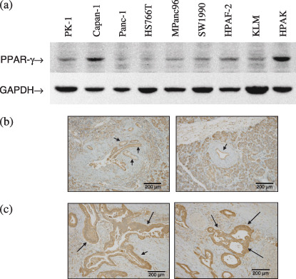
Expression of peroxisome proliferator‐activated receptor γ (PPARγ) in pancreatic cancer cells. (a) Western blots showing PPARγ expression in various pancreatic ductal adenocarcinoma cell lines, as well as a glyceraldehyde‐3‐phosphate dehydrogenase (GAPDH) internal control. Immunohistochemical staining of (b) normal ductal epithelium (left and right panels) and (c) pancreatic ductal adenocarcinoma (left and right panels) with anti‐PPARγ antibody. Arrows mark staining of normal pancreatic ductal epithelium in (b) and pancreatic ductal adenocarcinoma in (c).