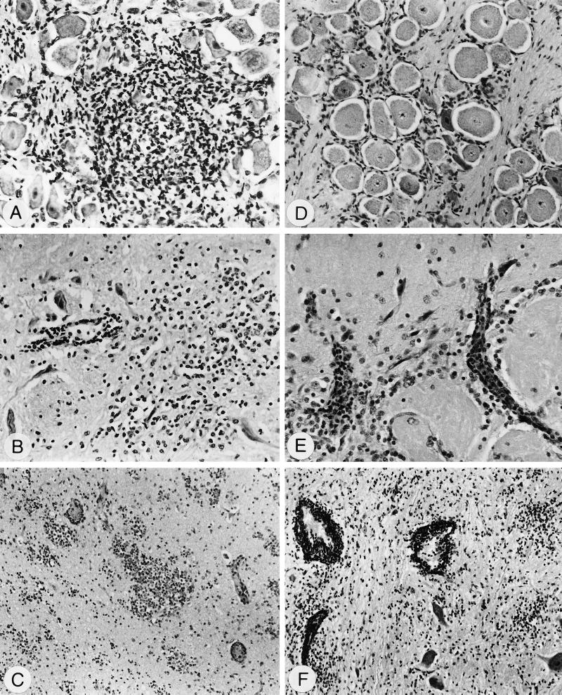
FIG. 5.
Histopathological alterations after infection with PrV-9112C2R (A to C) or PrV-9112C2 (D to F) visualized by hematoxylin-eosin staining. (A) Ganglion trigeminale, 2 dpi, ganglioneuritis, degeneration of perikarya, and focal infiltrations with macrophages and lymphocytes; magnification, ×200. (B) Medulla oblongata, 2 dpi, encephalitis, blood vessel with perivascular infiltrations (left) and prominent glia herds (right); ×200. (C) Bulbus olfactorius, 5 dpi, encephalitis, small vessels with perivascular infiltrates, and severe focal gliosis within the granular layer; ×100. (D) Ganglion trigeminale, 2 dpi, no inflammatory lesions at this time point; ×150. (E) B. olfactorius, 3 dpi, encephalitis, perivascular infiltrations within the glomerula olfactoria (bottom) and the molecular layer (top); ×240. (F) Pons, 6 dpi, encephalitis, severe perivascular infiltrates, and focal gliosis; ×100.

