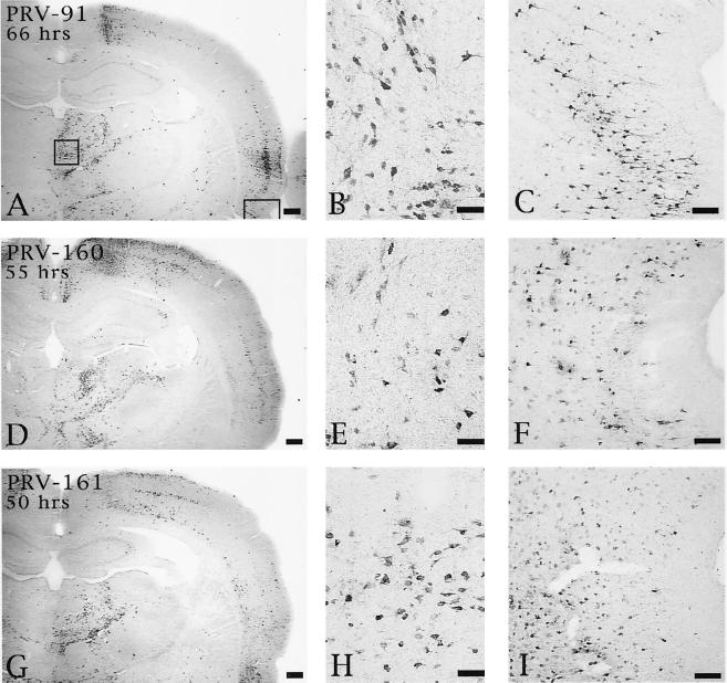
FIG. 7.
Extent of retrograde infection of thalamus and perirhinal cortex following injection of PRV 91 (A to C), PRV 160 (D to F), or PRV 161 (G to I) into PFC. The boxed areas shown in panel A designate the regions of the thalamus and perirhinal cortex shown at higher magnification in the photomicrographs to the right in each row (thalamus in panels B, E, and H; perirhinal cortex in panels C, F, and I). Note that the levels of retrograde infection produced by all three viruses are similar even though the animal infected with PRV 91 survived 66 h and those injected with PRV 160 and PRV 161 survived 55 and 50 h, respectively. Bars for A, D, and G, 500 μm; bars for B, E, and H, 50 μm; bars for C, F, and I, 100 μm.

