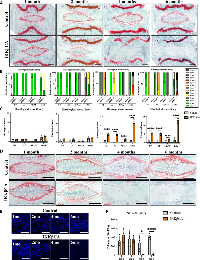
Fig. 4. IKKβ overexpression produces severe caudal DD.
(A) Representative images of safranin-O–stained mid-sagittal sections of control (AcanCre−/−;Ikk2cafl/fl) and IKKβCA caudal IVDs 1, 2, 4, and 6 months after recombination. Scale bars, 250 μm. Histological scoring legend, ranging from 0 (healthy) to 14 (most severe). (B) Distribution of histological scores. (C) Histological scoring within NP, AF, and NP:AF border compartments, and total score. (D) Representative images of safranin-O–stained mid-sagittal sections of the NP region of control and IKKβCA caudal IVDs at 1, 2, 4, and 6 months after recombination (n = 4 to 6 mice per genotype and time point, one to three caudal IVDs per mouse). Scale bars, 250 μm. (E) Representative images of 4′,6-diamidino-2-phenylindole (DAPI; nuclear)–stained mid-sagittal caudal IVD sections. Scale bars, 100 μm. (F) Quantification of caudal NP cellularity within hand-drawn ROIs of DAPI nuclear-stained mid-sagittal sections. *P < 0.05 and ****P < 0.0001. (n = 4 to 6 mice per genotype and time point, one to three caudal IVDs per mouse).

