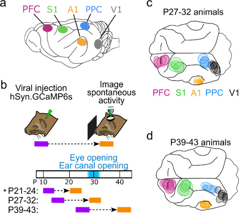
Figure 1: Imaging distinct cortical regions in ferrets at different developmental ages.

a. Target cortical locations. PFC – prefrontal cortex, S1 – somatosensory cortex, A1 – auditory cortex, PPC – posterior parietal cortex, V1 – visual cortex. b. Experimental timeline. Animals were injected with AAV expressing GCaMP6s 10–14 days prior to imaging. Imaging was performed at P27–32 or P39–43. P21–24 data from ref (Powell et al., 2024) (indicated by *) is presented for comparison with other ages. c. Imaged locations for P27–32 animals reconstructed from histology, colored based on assigned cortical area. d. Same as (c) for P39–43 animals.
