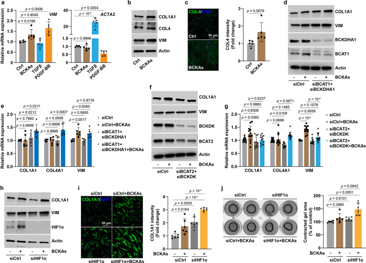
Fig 6. BCKAs promote a synthetic phenotype switch in PASMCs.
a, mRNA expression of synthetic marker VIM and contractile marker ACTA2 in BCKAs (100 μM), TGFβ (2 ng/mL), or PDGF-BB (10 ng/mL) stimulated cells. Fold change was relative to untreated controls. n = 5.
b, Representative immunoblots of synthetic marker proteins in BCKA-stimulated cells. n = 6.
c, Confocal microscopy images and quantitation represent COL4 synthesis and deposition in BCKA-treated PASMCs. Fold change was relative to control cells. n = 5.
d,e, Protein (d) and mRNA (e) expression of synthetic marker genes in PASMCs transfected with control siRNA (siCtrl) or human BCAT1 and BCKDHA1 siRNAs (siBCAT1+siBCKDHA1) with or without BCKA treatment. Fold change was calculated relative to siCtrl-transfected and untreated cells. n = 3 (d) and 6 (e).
f,g, Protein (f) and mRNA (g) expression of synthetic marker genes in PASMCs transfected with siCtrl or human BCAT2 and BCKDK siRNAs (siBCAT2+siBCKDK) with or without BCKA treatment.
Fold change was calculated relative to siCtrl-transfected and untreated cells. n = 3 (f) and 12 (g).
h, COL1A1 and VIM protein expression in control and HIF1α knockdown PASMCs in the presence or absence of BCKAs. n = 8.
i, Confocal microscopy and quantitation showing COL1A1 synthesis and deposition in control and HIF1α knockdown PASMCs in the presence or absence of BCKAs. Fold change was relative to control cells. n = 6.
j, Collagen gel contraction assay illustrating the relative contracted area of collagen gels after 8-hour of detachment in control and HIF1α knockdown PASMCs with or without BCKAs. n = 6.
All data are presented as mean ± SD. One-way ANOVA followed by Dunnett’s (a) or Tukey’s post-hoc test (e, g, i, j), or Kruskal-Wallis test followed by Dunn’s test (COL4A1 in g), or Mann-Whitney U test (c) was applied when compared to untreated controls (a, c), siCtrl-transfected and untreated or BCKA-treated PASMCs (e, g, i, j).

