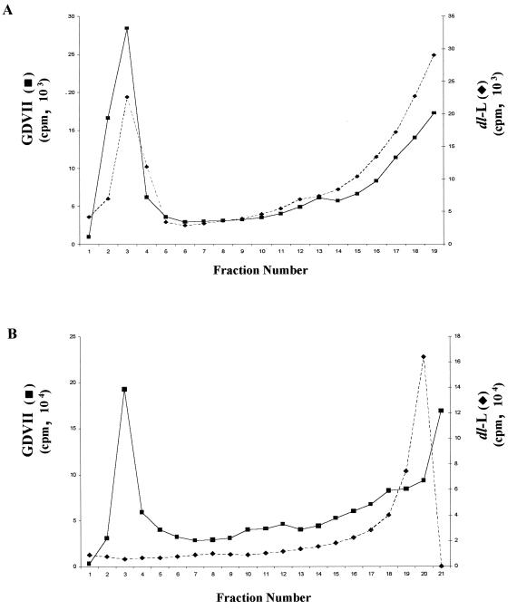
FIG. 7.
Virion assembly in BHK and L-929 cells. (A) BHK cells were infected with GDVII or dl-L virus stocks at an MOI of 5; then proteins were labeled with [35S]methionine from 5.5 to 8.5 h p.i. Lysates of infected cells were then fractionated through separate 15 to 30% sucrose gradients. (B) L-929 cells were infected with GDVII or dl-L virus stocks at an MOI of 80 and labeled from 5.5 to 8.5 h p.i.; then lysates were fractionated on 15 to 30% gradients. Fractions were immunoprecipitated, and then aliquots of each precipitate were counted in a scintillation counter. Intact virions sediment to the bottom of these gradients (fractions 1 to 5), and empty capsids migrate midway through the gradient (fractions 9 to 13).

