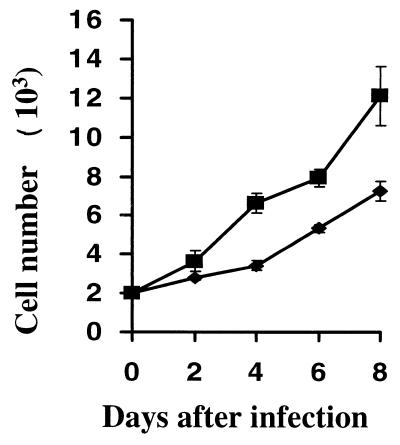
FIG. 5.
LMP1 increase the growth rate of MEF cells. MEF cells were freshly infected with v-LNSX (diamonds) or v-LMP1 (squares). After infection, the cells were removed from the plates with trypsin digestion at the end of P1; approximately 2,000 cells were plated in each well of 24-well plates in triplicate at day 0 and incubated further at 37°C. At the indicated time points, the cells were removed from the plates with trypsin and counted. The average numbers of cells at each time point was determined, and their standard derivations were obtained and plotted.

