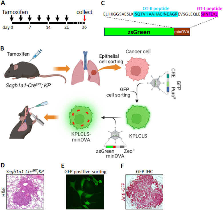
Figure 1. Generation of mouse lung adenocarcinoma cells.
(A) Schematic representation of the tamoxifen intraperitoneal injection schedule. Black arrowheads denote the time points at which tamoxifen was administered to each mouse. Lung tissues were harvested on day 36 for isolation of cancer cells. (B) Experimental workflow illustrating the overall process. Scgb1a1-CreERT; KP mice were injected with tamoxifen according to the schedule described above, followed by lung perfusion with digestion enzymes. Epithelial cells were isolated via fluorescence-activated cell sorting (FACS) and cultured. Isolated cells were transduced with a lentivirus bearing Cre to establish a stable cell line. Subsequently, a lentivirus carrying ZsGreen fused with minOVA was transduced into KPLCLS cells. Cancer cells were then engrafted intratracheally. (C) Amino acid sequence information of ZsGreen fused with minOVA. (D) Hematoxylin and eosin (H&E) staining of lung cancer induced by tamoxifen injections. (E) GFP+ cells were sorted by FACS and cultured in media supplemented with Puromycin. (F) KPLCLS-minOVA cells were inoculated via the intratracheal route and the lung tissues were collected after 36 days. Representative histological sections of lung cancer were stained with anti-GFP antibody (red).

