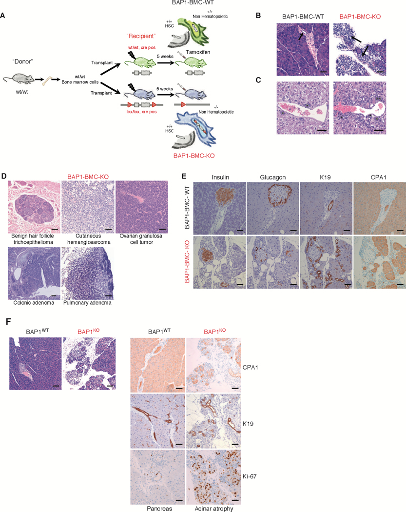
Fig. 1. A small proportion of mice with non-hematopoietic deletion of Bap1 develop diverse tumor types.

(A) Bone marrow cells from CD45.1+ wild-type mice were transplanted into lethally irradiated CD45.2+ Bap1+/+ creERT2+ (WT) and Bap1fl/fl creERT2+ (KO) recipients. Tamoxifen was given to recipients at 5 weeks after transplantation to induce Bap1 deletion. Mice were aged up to 10 months.
(B) Haematoxylin and eosin staining of pancreas 3 months post Bap1 deletion. Arrows indicate pancreatic ducts demonstrating expansion of duct profiles in the BAP1 deficient pancreas. Bar = 50 μM
(C) Haematoxylin and eosin staining of bile duct hyperplasia seen at 5–7 months post Bap1 deletion. Asterisks indicate bile ducts. Bar = 100 μM
(D) Haematoxylin and eosin staining of tumors seen at 10 months post Bap1 deletion.
Bar = 100 μM
(E) Immunohistochemical characterization of pancreatic acinar atrophy in 6-month-old BAP1 deficient mice; insulin, glucagon, carboxypeptidase A1 (CPA1), and keratin 19 (K19) immunolabeling. Bar = 50 μM
(F) Haematoxylin and eosin staining and immunohistochemical characterization of carboxypeptidase A1 (CPA1), keratin 19 (K19) and Ki-67 immunolabeling in the pancreas of mice with pancreatic specific BAP1deletion. Bar = 100 μM
