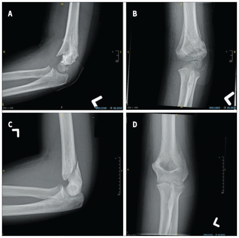
Fig. 2.
Radiographs of prospectively recruited patients who had management plan alterations because of postoperative radiographic assessment. Radiographs from patient 1 showed (A) malrotation at the fracture site and (B) varus alignment in the coronal plane at time of wire and immobilization removal. Radiographs from patient 2 showed (C) questionable bridging callus in the sagittal plane in a near skeletally mature patient with (D) an unstable fracture pattern.

