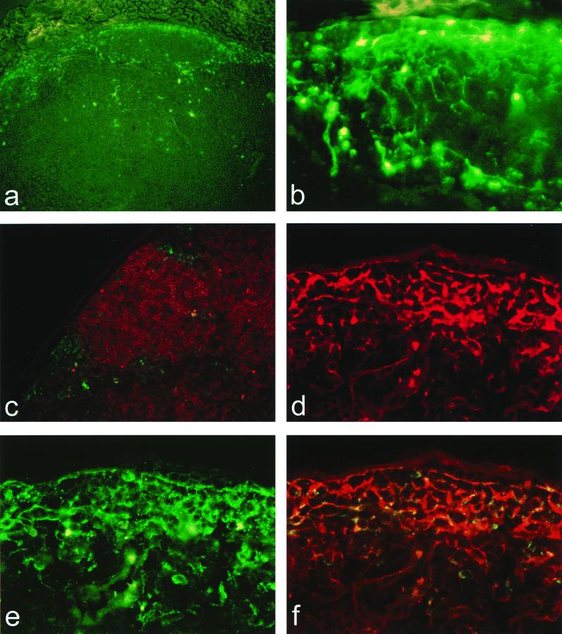
FIG. 2.
VEE infects DC in the draining lymph node. Sections of the draining popliteal lymph node 12 h following s.c. inoculation in the rear footpad either with 103 PFU of dpV3000-GFP (a to c), showing the distribution and morphology of GFP-positive cells in the cortex of the draining popliteal lymph node (a and b) (magnification, ×100 and ×400, respectively) and around B-cell follicles immunostained with the B-cell-specific Mab, B220 (c) (magnification, ×200), or with 103 PFU of V3000 (d to f), showing double immunostaining with VEE-specific Ab (CY2; green) and the DC-specific Ab, DEC 205, (Texas Red; red) visualized by using either Texas Red filters (d), fluorescein isothiocyanate filters (e), or triple-pass filters allowing simultaneous visualization of CY2 and Texas Red (f; magnification, ×400).

