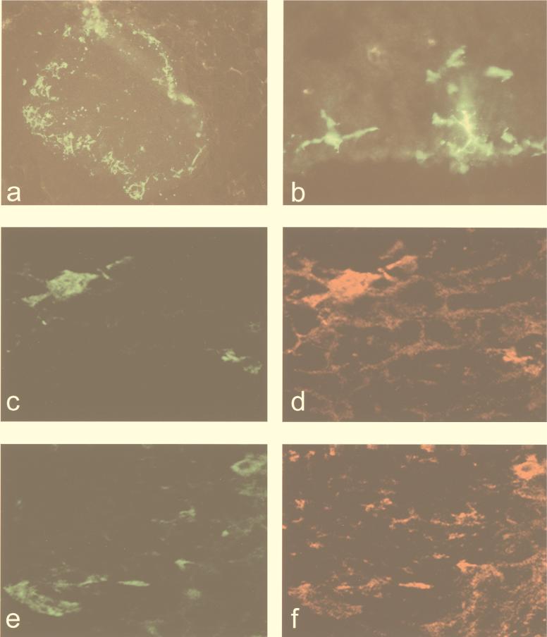
FIG. 3.
Langerhans cells are the first cells infected following inoculation. (a and b) A leg section showing the draining popliteal lymph node 12 h following s.c. inoculation in the rear footpad with 103 IU of GFP-VRP-V3000 (magnification, ×100 and ×400, respectively). (c to f) Draining lymph node sections from mice 8 h after being infected with 103 IU of HA-VRP-3000 were doubly immunostained with Ab specific for either influenza virus (c and e), DC (d; DEC 205), or MHC class II (f), and 0.5-μm-thick sections were analyzed by confocal microscopy (magnification, ×600).

