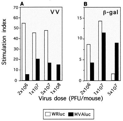
FIG. 6.
Specific Th cell proliferation after immunization of mice with WRluc or MVAluc virus. Splenocytes from mice of the same groups as used for the experiment depicted in Fig. 5 were tested for T-cell proliferation by stimulation in vitro with WR antigen (1 μg/ml; A) or β-Gal (1 μg/ml; B). After 72 h of culture, [3H]thymidine (1 μCi/well) was added; 18 h later, cells were harvested and radioactivity was measured. Bars represent the specific proliferative response measured as SI (cpm in the presence of the specific antigen/cpm in negative controls).

