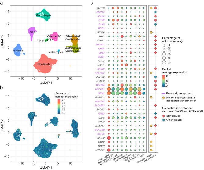
Fig. 3. Single-cell level gene expression patterns of CIE LAB values-associated genes.
a UMAP plot is shown for cell type identification. Cell types are identified with expression patterns of well-known cell type markers. Each color represents a cell type. b UMAP plot shows the overall expression patterns of CIE LAB-associated genes. Each cell is colored according to the average scaled expression of CIE LAB values-associated genes. c Dot plot of gene expression in cell types and additional evidence of each gene from the GWAS. Genes in purple represent those in previously unreported loci. The color of the circular dot represents the scaled average expression of each gene across cell types and size of the circular dot represents the percentage of cells expressing each gene within a particular cell type. Rhombic dots in yellow, red, and blue represent genes containing nonsynonymous variants associated with skin color, genes colocalized in skin tissue, or tissues other than skin, respectively. Abbreviations: Keratinocytes Diff. differentiated keratinocytes, Keratinocytes Undiff. undifferentiated keratinocytes, EC endothelial cells.

