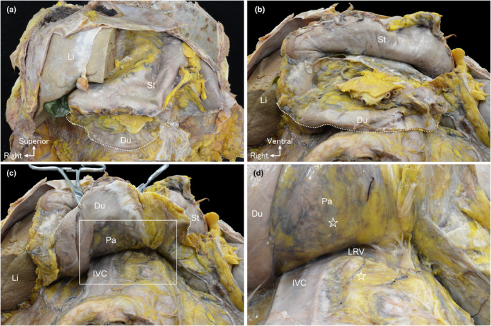
FIGURE 2.

Macroscopic anatomy of the duodenal mobilisation to the cranial side. (a) The ventral side of the upper abdomen following anterior abdominal wall removal. (b) The peritoneum at the inferior edge of the duodenum (dotted line) is incised. (c) The duodenum and the head of the pancreas are mobilised to the cranial side. (d) A magnified view of the white rectangular space in (c) reveals that mobilisation reaches the superior edge of the left renal vein (LRV). The dorsal surface of the head of the pancreas and the ventral surface of the retroperitoneal organs are covered with the fascia (☆). No evident nerves or vessels are observed penetrating through this fascia, visible to the naked eye between the superior border of the LRV and the inferior border of the duodenum. Du, duodenum; IVC, inferior vena cava; Li, liver; LRV, left renal vein; Pa, pancreas; St, stomach.
