
FIGURE 11.
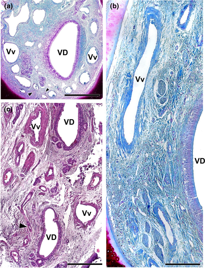
Vasculature of the wolf VNO. (a) Image of the anterior portion of the VNO is shown in Figure 8a exemplifies the presence of a profuse venous ring (Vv) surrounding the vomeronasal duct (VD). The arteries (arrowheads) are small and sparse. (b) Arteries indicated in A showed at higher magnification. (c) Caudal section of the VNO showing the glove‐fingered termination of the vomeronasal duct (VD). Numerous veins (Vv) and small arterial trunks (arrowhead) predominate at this level. (d) In a central section of the VNO, large venous sinuses (Vv) run along the lateral portion of the parenchyma. NCd, caudal nasal nerve. Staining: (a, b d) Gallego's trichrome; (c) haematoxylin–eosin. Scale bars: (a, d) = 250 μm; (c) = 100 μm; (b) = 50 μm.