
FIGURE 12.
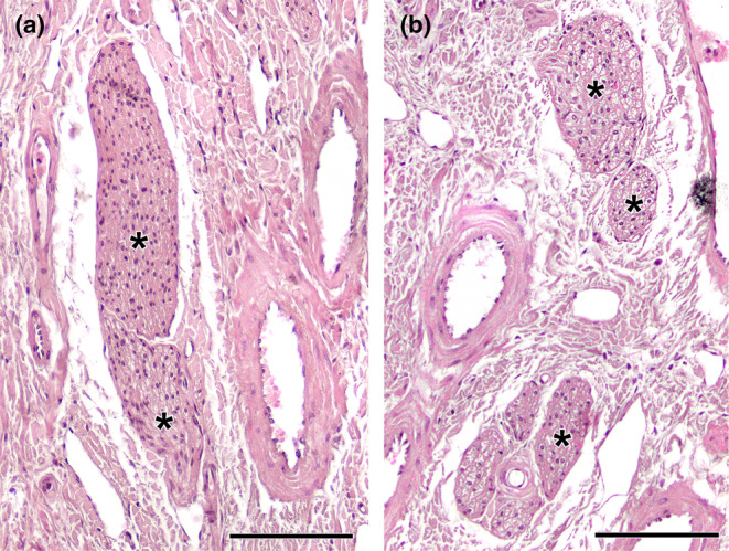
Histological study of the wolf vomeronasal organ innervation. (a). Unmyelinated branches of the vomeronasal nerves (*). They are characterised by their homogeneous appearance, with densely packed nerve bundles. (b). The lateral part of the parenchyma contains myelinated nerve branches originating from the caudal nasal nerve (*). Their structure is looser and the void spaces corresponding to the myelin sheaths are visible. Staining: Haematoxylin–eosin. Scale bar = 100 μm.