
FIGURE 16.
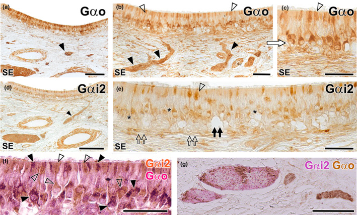
Immunohistochemical study of the wolf VNO. Gαo (a–c): Immunolabelling with anti‐Gαo shows a pattern of neuronal labelling predominantly concentrated in the neuroreceptor cells present in the basal layers of the neuroepithelium (a and b) and extending to the adjacent vomeronasal nerves (arrowheads). At higher magnification (c: enlarged area of the blue box in image b), it is appreciated how the labelling extends along the entire length of the immunopositive neuroreceptor cells, from the apical dendrite (open arrowhead) to the soma. Immunopositive neuroreceptor cells embrace the intraepithelial capillaries of the VNO. Gαi2 (d, e): The labelling is predominantly concentrated in the neuroreceptor cells mainly located in the central zone of the epithelium (asterisk) and the vomeronasal nerves (arrowhead). Unlike Gαo, no immunopositive neurons are identified around the intraepithelial capillaries (black double arrow). The dendritic knobs are less numerous than in Gαo but more dilated (open arrowhead). The deep neuronal clusters are immunonegative (open double arrows). (f–g): f Double immunostaining for Gαi2 and Gαo confirms the presence of two subpopulations of vomeronasal neuroreceptor cells. Anti‐Gαi2 (brown) immunostains a subpopulation of cells predominantly located in the central zones, which have thick dendritic knobs (open arrowheads). Anti‐Gαo (red) stains a cell subpopulation mainly located in a more basal zone (black arrowheads). (g) In some cases, as the one shown, the vomeronasal nerves in the parenchyma are predominantly composed either of Gαi2 type fibres (red) or Gαo type fibres (brown). Scale bars: (a, d and g) = 100 μm; (b, c, e and f) = 50 μm.