
FIGURE 19.
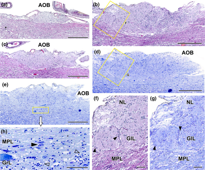
Histological study of the wolf accessory olfactory bulb stained with haematoxylin–eosin and Nissl staining. (a–c). A general view of the AOB can be seen at three selected sagittal levels stained with HE. They show the elongated shape of this structure and the predominance of the nervous (NL) and glomerular layers (GlL). (d, e) Sagittal Nissl‐stained sections at low magnification allows to appreciate the development of the AOB. (f) At higher magnification (corresponding to box in b), two glomerular formations clearly defined by periglomerular cells (arrowheads) are appreciated. (g) The magnification of the superficial area of the AOB (box in d) allows to discriminate the presence of a mitral‐plexiform layer (MPL). (h) Enlargement of the deep area of the AOB (corresponding to box in e) shows mitral cells (black arrowhead) in the MPL as well as granular cells (open arrowhead) in the deeper granular layer (GrL). Scale bars: (a–e) = 500 μm; (f–h) = 100 μm.