
FIGURE 20.
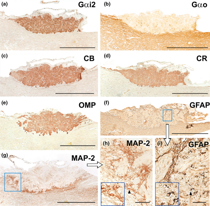
Immunohistochemical study of wolf AOB. (a) Anti‐Gαi2 uniformly and intensely label the superficial layers of the AOB (nervous and glomerular layers). The entire surrounding neuropil is negative. (b) Anti‐Gαo produces a reverse pattern to the one shown in A, where the neuropil surrounding the superficial layers is strongly immunopositive, including the mitral‐plexiform and granular layers of the AOB. However, the superficial layer is clearly negative, although immunopositive punctae areas are observed. (c–e) The calcium binding proteins, calbindin (c) and calretinin (d), as well as OMP (e) show an identical labelling pattern to that obtained with anti‐Gαi2, concentrated in both the nerve and glomerular layers and immunonegative for the neuropil. (f) Anti‐GFAP (f and enlarged area in i) produces a trabecular labelling pattern in the nerve and glomerular layers, which corresponds to the ensheathing glia accompanying the vomeronasal nerve endings. Occasionally, cell bodies belonging to these glial cells are visible (arrowhead). The cell body is magnified in the bottom left‐hand box. (g) Anti‐MAP‐2 immunolabelling does not produce immunopositive labelling in the superficial layers (nervous and glomerular), but it strongly labels an irregular band corresponding to the MPL layer. (h) MAP‐2‐immunopositive prolongations originating from the MPL can be observed running between the glomeruli of the AOB. (h: enlargement of the box in g). The cell body of an immunopositive interneuron (arrowhead) is shown in an enlarged view in the box at the bottom left. Scale bars: (a–g) = 500 μm; (h–i) = 50 μm.