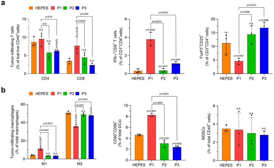
Extended Fig. 4. P1 promotes innate and adaptive immune activation within the tumor microenvironment.
(a) P1 increased the population of tumor-infiltrating CD8+ T cells and IFNγ-producing CD8+ T cells, but decreased that of Tregs (CD3+CD4+CD25+FoxP3+), relative to P2 and P3 (n=3, mean±SD). Tumor-infiltrating lymphocytes were obtained on day 16 (2 days after the last treatment). (b) P1 promoted M1 macrophage polarization (M1 macrophage: CD206−CD80+CD86+; M2 macrophage: CD206+CD80−; macrophages: CD11b+CD11c−F4/80+MHC-II−) and maturation of dendritic cells (DCs) (Mature DCs: CD80+CD86+ DC; DC: CD11c+MHC-II+F4/80−) but did not affect the number of myeloid-derived suppressor cells (MDSCs) (CD11b+CD11c−MHC-II−F4/80−Gr-1+) within the tumor microenvironment relative to P2 and P3 (n=3, mean±SD), unpaired Student’s t test in comparison with Cont and the indicated conditions.

