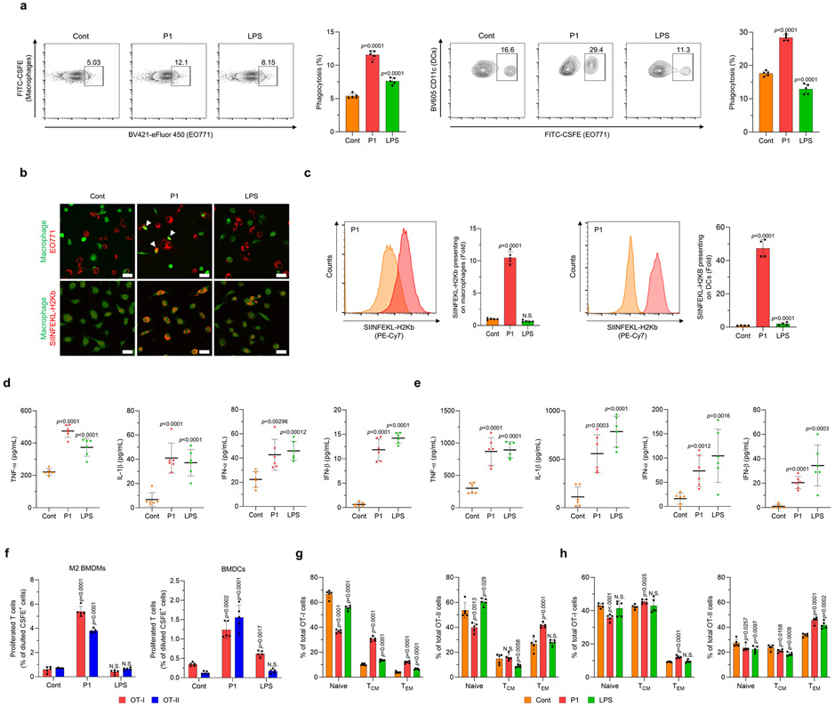
Fig. 4. P1 enhances the phagocytosis of cancer cells and the priming of antigen-specific T cells by professional antigen-presenting cells.
(a) P1 increased the phagocytic activity of M2 bone marrow-derived macrophages BMDMs and BMDCs, as evaluated by flow cytometry (n=5, mean±SD). (b) Macrophages promoted the phagocytosis of EO771 cells (white arrows) and cross-presentation of ovalbumin (OVA) peptide (SIINFEKL-H2Kb) on their surface, as visualized by confocal laser scanning microscopy; scale bar 20 μm. (c) P1 promoted cross-presentation of SIINFEKL-H2Kb peptides on the surfaces of M2 BMDMs (n=5, mean±SD) and BMDCs (n=4, mean±SD), as assessed by flow cytometry to normalize mean fluorescence intensity of SIINFEKL-H2Kb peptides; unpaired Student’s t test in comparison to Cont. (d,e) P1 increased the production of pro-inflammatory cytokines (TNF-α, IL-1β) and type I interferons (IFN-α and IFN-β) in co-cultures of EO771 cancer cells with (d) M2 BMDMs or (e) BMDCs (n=6, mean±SD). (f) P1 induced the proliferation of both CD4+ and CD8+ T cells isolated from transgenic OT-I and OT-II mice and co-cultured with P1-treated M2 BMDMs or BMDCs and cancer cells (n=5, mean±SD). (g,h) P1 activated T central memory (TCM) and T effector memory (TEM) subtypes of both CD4+ and CD8+ T cells with (g) M2 BMDMs or (h) BMDCs (n=5, mean±SD); unpaired Student’s t test in comparison to Cont.

