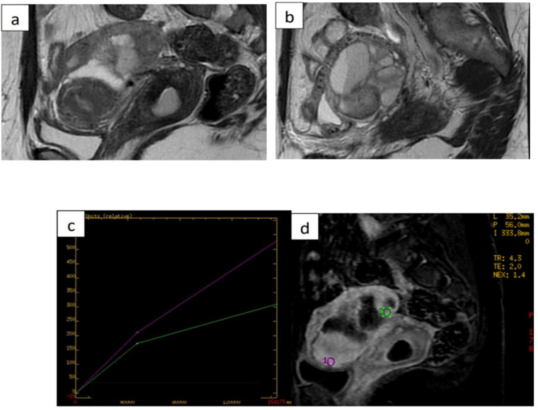
Figure 2.
Images from 38-Year-Old Woman with Left-Sided Cystic Adnexal Mass Indeterminate in Transvaginal Ultrasound. (a) and (b) sagittal T2-weighted magnetic resonance (MR) images show leftsided multiloculated adnexal mass, with largest diameter of 52mm, with intermediate signalintensity locules and intermediate-signal-intensity thickened and irregular internal septa. (c) and (d) dynamic contrast enhanced MRI sequence yielded intermediate time-intensity curve with type-2 plateau (green line) in comparison with adjacent external myometrium (purple line). Histology confirmed benign infected endometrioma. ADNEX MR score was upgraded to 4 (type-2 curve)

