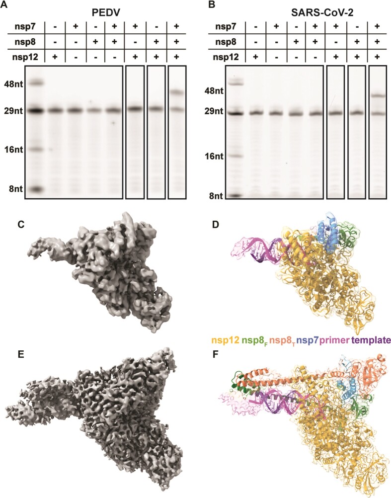
Figure 1.
Assembly of an active PEDV polymerase complex. A 29 nt RNA primer with a 5′ fluorophore is annealed to a 38 nt template and extended in the presence of CoV polymerase complexes. Combinations of nsp7, nsp8, and nsp12 were tested for PEDV (A) and SARS-CoV-2 (B). (C, E) 3.3 and 3.4 Å cryo-EM reconstruction of the PEDV core polymerase complex with (E) and without (C) nsp8T, respectively. (D, F) Coordinate models of the PEDV core polymerase complexes docked into their corresponding electron density maps colored by chain.

