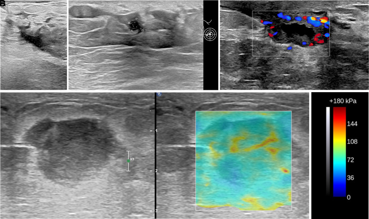
Figure 2. A-E.
Targeted ultrasound images from (A) through (D) depicting different grades of idiopathic granulomatous mastitis (IGM) on different patients. (A) A fistula tract shows the drainage of purulent content on the background B-mode image and color Doppler map displays ongoing hyperemia within the wall of the tract. This finding corresponds to Grade II. (B) Hypoechoic solid-like mass measuring about 3.4 cm on long axis with diffuse increment on echogenicity of surrounding tissue obtained at gray-scale ultrasound at 9 o’clock position of the right breast in a 28-year-old postpartum woman presenting with denial of breastfeeding by her baby on the affected breast. Following 3 weeks of conservative treatment due to a lack of symptom resolution with progression in the size of the lesion, a biopsy was performed, and the result was IGM. This lesion demonstrates the earliest course of disease initiation and corresponds to grade I. (C) In another patient with a history of periductal mastitis on the contralateral breast prior 2.5 years was referred to the tertiary university hospital for further work-up of the lesion on the right side. The abscess formation (measurement markers) is clearly depicted within the extremely IGM-involved periareolar breast side. These findings correspond to grade III. (D) Elastosonographic image of the biopsy-proven IGM lesion. Color box (E) indicates the stiffness of tissue based on elasticity properties of the region of interest with red depicting the hardest and blue the softest.

 Content of this journal is licensed under a