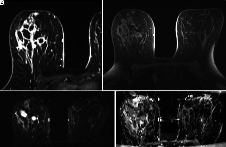
Figure 3. A-D.
Idiopathic granulomatous mastitis (IGM) in a 24-year-old patient with a persistent history of IGM on the right breast for the past 3.2 years. A thorough evaluation was not yielding for any etiologic agent. The lesions on magnetic resonance imaging (MRI) show extensive segmental involvement with diffuse contrast enhancement on (A) flow dynamic and (B) post-contrast subtraction images. The mass shows restricted diffusion on (C), axially acquired image from diffusion-weighted MRI (b = 800 s/mm2) and a low apparent diffusion coefficient on (D). The patient underwent 5 courses of intralesional steroid injection therapy and no relapse was noted on 3 years of follow-up.

 Content of this journal is licensed under a