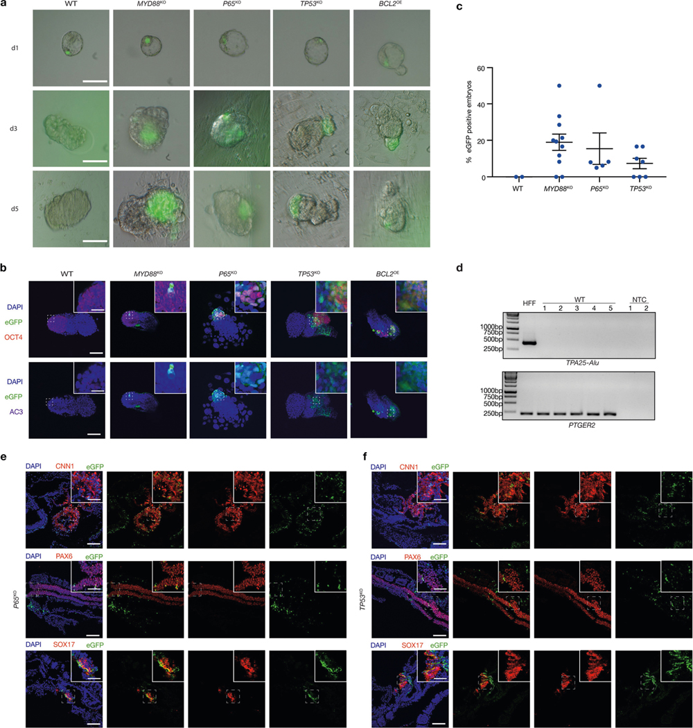
Extended Data Fig. 8 |. Overcoming interspecies PSC competition enhances survival and chimerism of human primed PSCs in early mouse embryos.

a, Representative brightfield and fluorescence merged images of mouse embryos cultured for 1 day (d1), 3 days (d3) and 5 days (d5) after blastocyst injection with wild-type, MYD88KO, P65KO, TP53KO and BCL2OE hiPS cells. Scale bars, 100 μm. b, Representative immunofluorescence images of day-5 mouse embryos co-stained with OCT4 (red), eGFP (green) and AC3 (purple) after blastocyst injection with wild-type, MYD88KO, P65KO, TP53KO and BCL2OE hiPS cells. Top, eGFP and OCT4 merged images with DAPI; bottom, eGFP and AC3 merged images with DAPI. Scale bars, 100 μm and 50 μm (insets). c, Dot plot showing the percentages of eGFP+ E8–E9 mouse embryos derived from wild-type, MYD88KO, P65KO and TP53KO hiPS cells. Each blue dot represents one embryo transfer experiment. n = 2 (WT), n = 11 (MYD88KO), n = 5 (P65KO) and n = 7 (TP53KO), independent experiments (Supplementary Table 2). d, Genomic PCR analysis of E8–E9 mouse embryos derived from blastocyst injection of wild-type hiPS cells. TPA25-Alu denotes a human-specific primer. PTGER2 was used as a loading control. HFF, HFF-hiPS cells. NTC, non-template control. The experiment was repeated independently three times with similar results. For gel source data, see Supplementary Fig. 1. e, f, Representative immunofluorescence images showing contribution and differentiation of eGFP-labelled P65KO (e) and TP53KO (f) hiPS cells in E8–E9 mouse embryos. Embryo sections were stained with antibodies against eGFP and lineage markers including CNN1 (mesoderm, top), PAX6 (ectoderm, middle) and SOX17 (endoderm, bottom). Scale bars, 100 μm and 50 μm (insets). Images in a, b, e and f are representative of three independent experiments.
